What is a fractured endodontic instrument?
Endodontic files are the small, precision instruments used to clean and shape root canals during treatment. The majority of modern rotary files are made from nickel-titanium (NiTi) alloy — a material valued for its flexibility and cutting efficiency in curved canals. However, NiTi files can fracture inside the canal under certain conditions, leaving a metallic fragment that can block access to the remainder of the root canal system.
Instrument fracture is a recognised complication of root canal treatment — it can occur even with careful technique, high-quality instruments, and experienced operators. Fracture is more common in severely curved canals, heavily calcified canal systems, canals with unexpected anatomy, and where files have been used repeatedly. It is not automatically a reflection of poor treatment or operator error. That said, when it does occur, appropriate management is essential.
Is a fractured instrument always a problem?
Not necessarily. The significance of a fractured instrument depends on several factors, and a calm, systematic assessment is always the right first step.
Prognosis is often good even without retrieval when:
- The canal was adequately cleaned before the fracture occurred
- There is no periapical pathology associated with the tooth
- The tooth is asymptomatic
- The fragment is in a clean, well-obturated portion of the canal
Prognosis is more guarded and active management is required when:
- The fragment is blocking access to an infected or incompletely cleaned portion of the canal
- The tooth is symptomatic — pain, swelling, sinus tract
- Periapical pathology is present on radiographic examination
CBCT 3D imaging is frequently invaluable in assessing the exact position of a fractured fragment and determining the most appropriate management strategy.
When should a fragment be removed versus left in place?
The decision to attempt retrieval, bypass, or monitor is based on an honest assessment of the clinical situation — not a blanket policy of always removing every fragment.
Retrieval is generally indicated when:
- The fragment is blocking an infected or uncleaned portion of the canal
- The tooth is symptomatic with pain, swelling, or a persistent sinus tract
- Periapical pathology is present and attributable to the inadequately treated canal
Monitoring may be appropriate when:
- The fragment is in a well-cleaned canal segment with no periapical pathology
- The tooth is asymptomatic and the canal below the fragment has been adequately treated
- The retrieval risk is disproportionate to the likely benefit — for example, a fragment deep in a severely curved canal
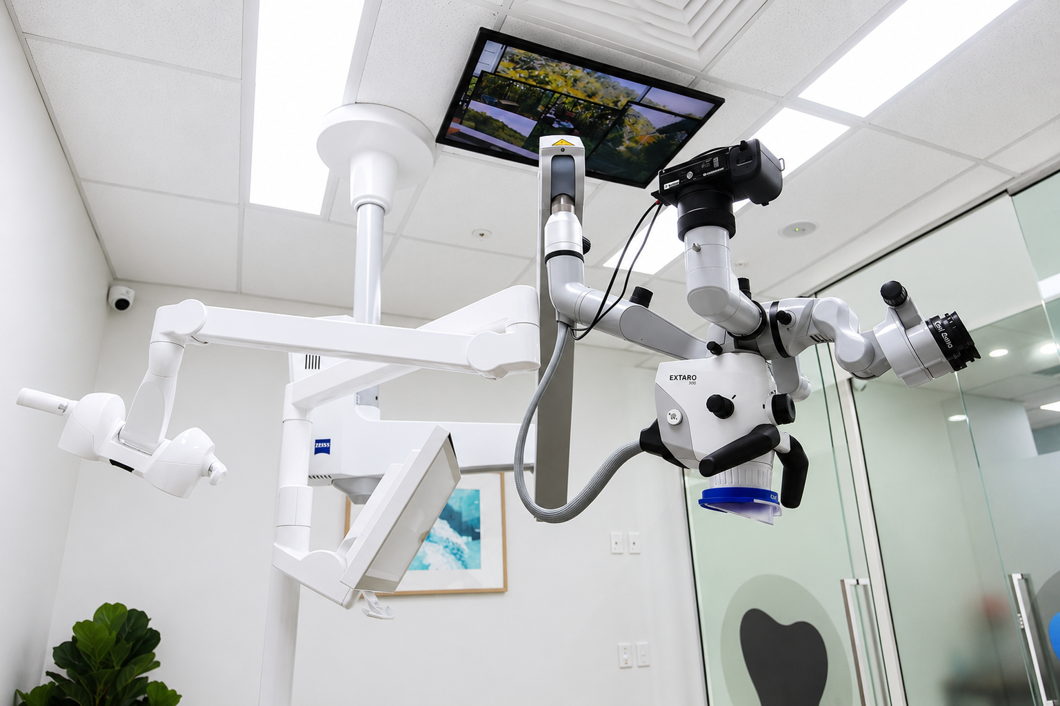
Our approach — operating microscope and ultrasonic retrieval
The operating microscope is not optional for fractured instrument management — it is essential. Fragments are often invisible to the naked eye and can only be precisely located and accessed under high magnification.
Under the microscope, specialised ultrasonic tips are used to vibrate around the coronal portion of the fragment, creating a trough in the surrounding dentine. This loosens the fragment's purchase within the canal walls, allowing it to be retrieved with dedicated extraction instruments or to be flushed coronally with irrigation.
The success of retrieval depends primarily on:
- Fragment position: coronal and middle third fragments are generally more accessible than apical third fragments
- Canal curvature at the fracture site: a fragment in a straight section of canal is far more retrievable than one at the apex of a severe curve
- Fragment length: longer fragments provide more surface area for the ultrasonic tip to act on
We are always honest about the limitations of retrieval. We will not attempt removal where the risk of causing further damage — including dentine removal, ledging, or perforation — outweighs the potential benefit.
When bypassing or surgery is preferred
In many cases, bypassing the fragment — negotiating past it with a fine hand file — is preferable to aggressive retrieval attempts. When bypass is achieved, the canal can be cleaned and filled to the working length with the fragment incorporated into the obturation. This avoids the risks associated with repeated ultrasonic activation and dentine removal during retrieval attempts.
Where a fragment cannot be retrieved or bypassed and the tooth remains symptomatic with periapical pathology, apical surgery may be the most appropriate management — directly addressing the periapical lesion without requiring access through the fragment.
Referring your patient
To ensure we can assess your patient promptly and accurately, please include in your referral:
- The specific tooth and canal in which the fracture occurred
- Estimated position of the fragment — coronal, middle, or apical third
- Pre-operative and post-operative periapical X-rays
- Current clinical status — whether the tooth is symptomatic
- Details of any previous retrieval attempts
Submit your referral via our online referral form or call us on (02) 9129 8806 to discuss the case before referring.
Refer for fractured instrument assessment
Call us to discuss retrievability before you refer — we are happy to advise on the risk-benefit profile for your specific case.
Frequently asked questions
Can a fractured instrument always be removed?
No — not every fractured instrument can be retrieved. Success depends on the position of the fragment in the canal, the degree of canal curvature at the fracture site, and the length of the fragment. Fragments in the coronal or middle thirds of straight canals are often retrievable. Fragments deep in the apical third of a curved canal are significantly harder to remove, and attempts carry a risk of further complications. We discuss the risk-benefit profile honestly before any attempt is made.
What happens if the instrument cannot be removed?
If retrieval is not possible or not advisable, there are other management options. Bypassing the fragment — negotiating past it with a smaller file — can allow completion of canal cleaning and filling even with the fragment in place. In cases where the tooth remains symptomatic and the fragment cannot be bypassed, apical surgery may be indicated to address the periapical pathology directly. In some situations, monitored observation is appropriate.
Does a fractured instrument mean my root canal will fail?
Not necessarily. The prognosis depends primarily on whether the canal was adequately cleaned before the fracture occurred, and whether infection is present. If a file fractures in a well-cleaned, uninfected canal, the long-term prognosis may be good even if the fragment cannot be removed. If the fracture occurred before adequate cleaning was completed and there is periapical infection, the prognosis is more guarded and active management is usually required.
How long does instrument retrieval take?
Instrument retrieval is a time-consuming and technically demanding procedure. Most retrieval appointments are scheduled for 60–90 minutes. In some cases a second appointment is needed. The time required depends on the position and accessibility of the fragment, the degree of canal curvature, and whether bypassing is needed as an adjunct to retrieval.
When should I refer a patient with a separated instrument?
Refer promptly if the tooth is symptomatic, if there is periapical pathology present, or if the fragment is blocking an uncleaned or infected portion of the canal. If the fragment occurred in a well-cleaned canal with no signs of infection, a period of monitoring may be appropriate — but a specialist opinion is always worthwhile. Include the affected tooth and canal, estimated fragment position, periapical X-rays, and any previous retrieval attempts in your referral.